Radiographic Interpretation for Dental Hygienists
Sharpen image assessment skills to identify calculus, bone loss, and pathology and support the diagnostic workflow.
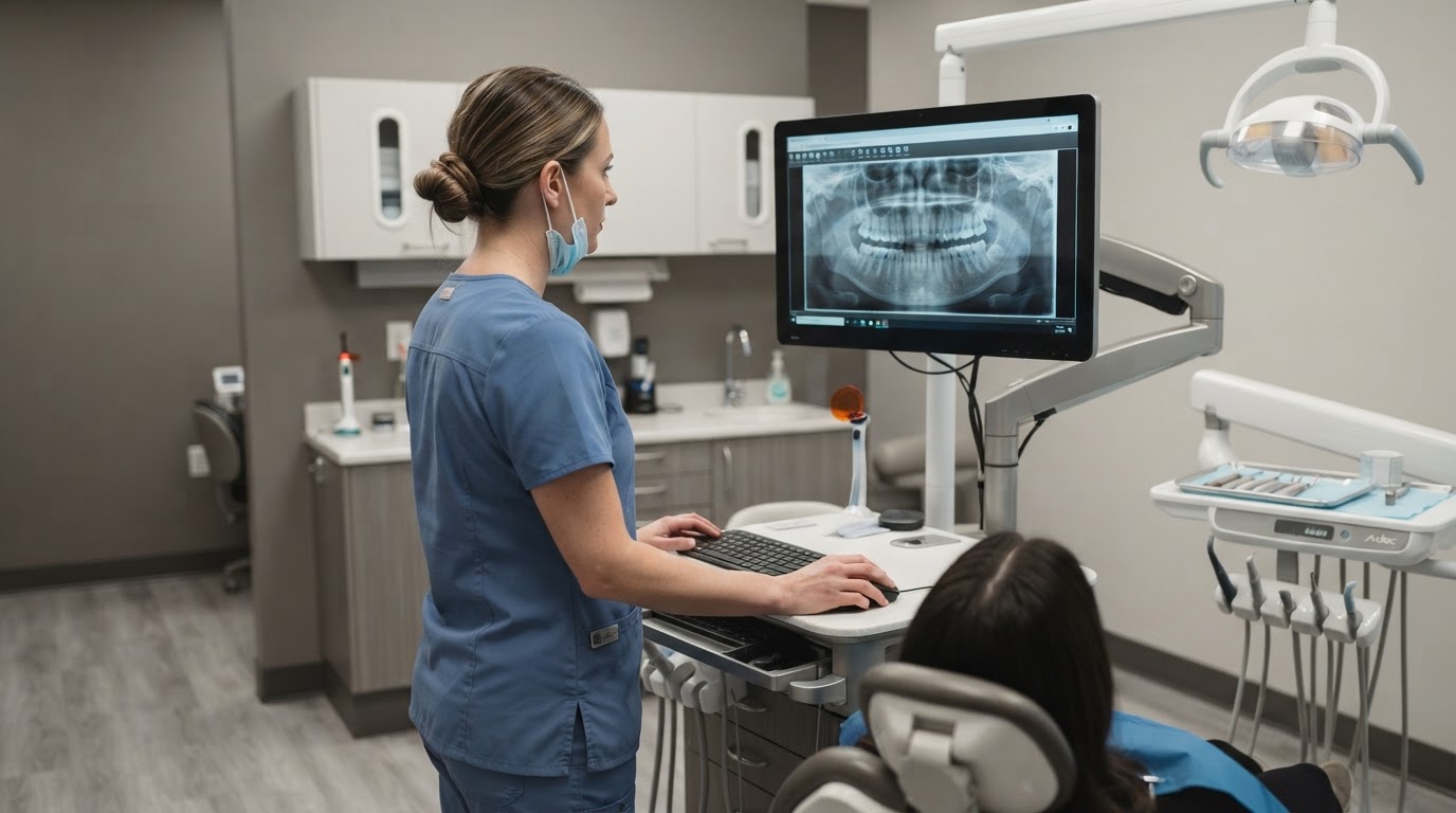
The Hygienist's Role in Assessment
Dental hygienists assess radiographs to identify findings relevant to the hygiene appointment, including calculus, altered bone levels, and obvious pathology. Definitive diagnosis remains with the dentist, but a sharp assessment accelerates the workflow and improves patient communication.
A Systematic Reading Pattern
- Read every image in the same sequence to avoid missed findings.
- Evaluate crestal bone height relative to the cementoenamel junction.
- Look for spiky, burnished interproximal calculus shadows.
- Compare the horizontal and vertical pattern of any bone loss.
Correlating With the Clinical Exam
Radiographs are a two-dimensional summary. Always correlate radiographic findings with the periodontal chart, especially for furcation involvement and early bone loss that may not yet be visible on film.
Practical Takeaway
Use a consistent reading sequence on every image, correlate findings with the periodontal chart, and remember that radiographs underrepresent both early bone loss and burnished subgingival calculus.
Authoritative Dental Resources
Join the RDHPlanet Weekly Newsletter
Practical, evidence-aware insights for working dental hygienists. Delivered straight to your inbox every Sunday.
Related Articles
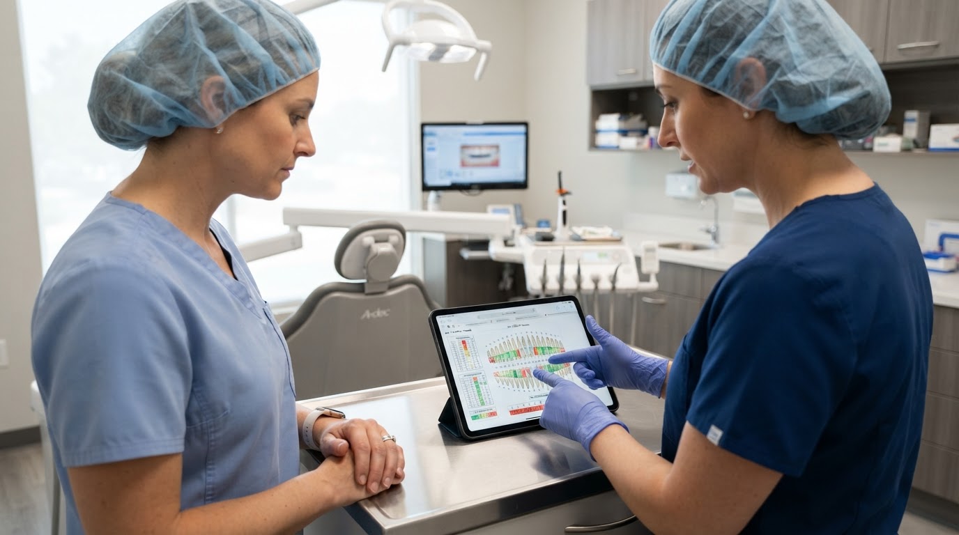
Periodontal Charting: A Reliable Assessment Protocol
A standardized probing protocol improves diagnostic accuracy and supports defensible, reproducible periodontal records.
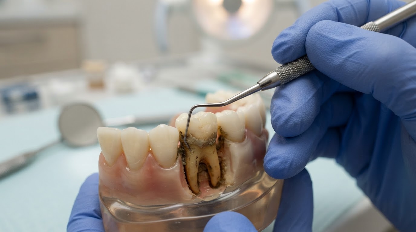
Calculus Detection Techniques That Improve Outcomes
Combine tactile, visual, and adjunctive detection methods to find subgingival calculus you might otherwise miss.